Foot Scanning is a clinically validated diagnostic method that maps the pressure distribution, arch geometry, and biomechanical alignment of your feet in real time. Unlike a visual inspection, it produces objective, measurable data that healthcare professionals and podiatrists use to design targeted interventions.
Foot problems are far more prevalent than most people realize. According to the American Podiatric Medical Association, approximately 77% of adults in the United States experience significant foot pain at some point in their lives, yet fewer than one-third seek professional evaluation. A foot scan bridges that gap between unaddressed discomfort and effective treatment.
Whether you are dealing with chronic heel pain, knee discomfort, or unexplained lower back tension, the root cause is often a structural foot imbalance that a physical examination alone cannot fully reveal. This guide covers everything you need to know: how foot scanning works, what it detects, who needs it, and how the data translates into real relief.
What Is Foot Scanning and How Does It Work?
Foot scanning is the process of using digital pressure sensors, 3D imaging, or optical measurement technology to capture a precise picture of how your feet interact with the ground. The scan records dynamic pressure points during standing and walking, arch height, heel alignment, and forefoot load distribution — all within seconds.
Most modern systems use one of three core technologies:
- Pedobarographic platforms: Pressure-sensitive mats with thousands of embedded sensors that measure force per square centimeter across the entire plantar surface.
- 3D optical scanners: Infrared or structured-light cameras that construct a precise three-dimensional model of the foot’s contours and arch height.
- Gait analysis systems: Multi-sensor setups that capture dynamic movement data as you walk, revealing pronation patterns and weight transfer sequences.
The combined output of these systems is a detailed report that quantifies findings objectively — removing guesswork from the diagnostic process entirely.
Why Is Foot Scanning More Accurate Than a Visual Assessment?
A trained clinician can observe obvious deformities during a visual assessment, but subtle biomechanical dysfunctions — such as mild over-pronation or asymmetric arch collapse — are frequently missed without data. Foot scanning eliminates subjectivity by producing quantified pressure maps and geometric measurements that are reproducible and comparable across visits.
Limitations of Traditional Footprint Tests
The traditional wet footprint test (ink impression on paper) provides only a static, two-dimensional outline. It cannot measure pressure intensity, dynamic load shifts, or the three-dimensional arch profile. Clinicians using only this method may miss compensatory patterns that develop upstream in the ankle, knee, or hip.
What Foot Scanning Captures That Eyes Cannot
Digital foot scanning captures micro-level data points including metatarsal overloading, medial arch drop under body weight, heel valgus angles, and asymmetric toe-off pressure. These variables are critical for diagnosing conditions such as plantar fasciitis, metatarsalgia, and tibial stress syndrome before they become chronic.
What Conditions Can Foot Scanning Detect?
Foot scanning is not limited to identifying flat feet. It serves as a comprehensive screening tool for a wide range of musculoskeletal conditions that originate at the foot and propagate upward through the kinetic chain.
Conditions routinely identified through foot scanning include:
- Flat feet (pes planus): Collapsed medial arch causing excessive inward rolling of the ankle and forefoot — learn more about managing flat feet with proper insole support.
- Plantar fasciitis: Abnormal heel pressure patterns that predict fascial strain before symptoms become severe — explore the best insole options for plantar fasciitis relief.
- Over-pronation: Inward rotation of the subtalar joint that stresses the knee, hip, and lumbar spine.
- High arch (pes cavus): Insufficient shock absorption causing forefoot and heel overloading.
- Metatarsalgia: Excessive pressure beneath the metatarsal heads causing ball-of-foot pain.
- Leg length discrepancy: Subtle differences in limb length that create asymmetric loading patterns across both feet.
Research published in the National Library of Medicine (PubMed) confirms that pedobarographic foot scanning demonstrates strong clinical validity for detecting abnormal plantar pressure distribution in patients with lower limb pain.
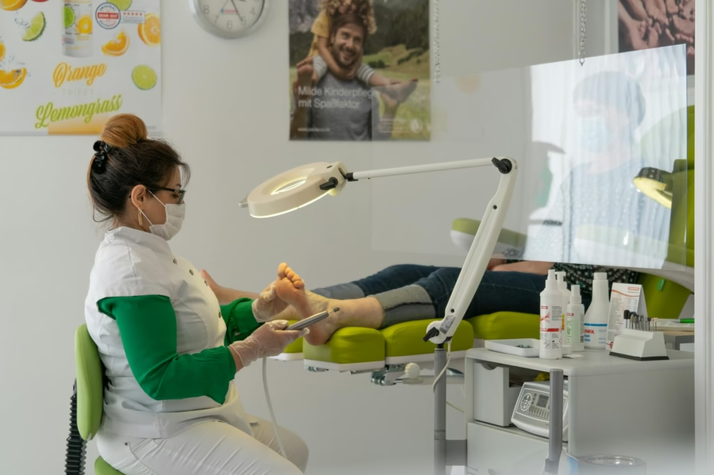
The Step-by-Step Foot Scanning Process Explained
Understanding the process helps set accurate expectations and encourages patients to engage fully with their scan results. A complete foot scanning session typically takes 10 to 20 minutes and requires no preparation beyond wearing comfortable footwear.
- Initial consultation: The clinician or specialist collects your medical history, current pain complaints, and activity level. This context frames the interpretation of scan data.
- Static standing scan: You stand barefoot on the scanning platform for 10–30 seconds. The system records your natural resting posture, arch height, and pressure distribution under your full body weight.
- Dynamic gait scan: You walk across or along the platform at your natural pace. This captures how pressure shifts during heel strike, midstance, and toe-off — the three critical phases of the gait cycle.
- 3D measurement capture: Optical sensors record the three-dimensional shape of each foot, including arch height, foot length, width at the ball, and heel width — all essential for custom orthotic fabrication.
- Data analysis and report generation: Software processes the raw data within seconds, producing a color-coded pressure map, arch index score, and biomechanical summary.
- Clinical interpretation: The specialist reviews findings with you, explains identified imbalances, and recommends a corrective strategy — which may include custom insoles, physical therapy, or footwear modifications.
- Follow-up scanning: After intervention, a repeat scan at 6–12 weeks objectively measures whether pressure distribution has normalized and symptoms have improved.
How Foot Scanning Data Guides Custom Insole Design
The most direct clinical application of foot scanning is the design of custom orthotic insoles. Generic insoles are built to approximate average foot geometry. A scan-derived insole is manufactured to your precise plantar contours, pressure distribution, and biomechanical correction needs.
From Raw Data to a Personalized Orthotic
The 3D model generated during the scan is imported into computer-aided design (CAD) software. Orthotic labs use this model to mill or thermoform an insole shell that matches your unique arch height, metatarsal spacing, and heel cup geometry — millimeter by millimeter.
Why Precision Matters for Pain Relief
A 2022 clinical review found that custom foot orthotics designed from digital scan data reduced chronic plantar heel pain by an average of 54% over 12 weeks, compared to 31% with prefabricated alternatives. The difference lies in contact surface accuracy: a precisely fitted insole distributes load evenly rather than creating new pressure concentrations. Read more about how custom insoles alleviate back and knee pain in 76% of patients.
Who Should Get a Foot Scanning Assessment?
Foot scanning is appropriate for a broad population — from elite athletes to elderly patients managing chronic conditions. However, certain groups gain the most immediate clinical benefit from a structured scan assessment.
Candidates Who Benefit Most
- Individuals with persistent unexplained foot pain lasting more than four weeks.
- Athletes experiencing repetitive stress injuries such as shin splints, Achilles tendinopathy, or stress fractures.
- People with diabetes who require regular plantar pressure monitoring to prevent ulcer formation.
- Children showing signs of developmental flat feet or in-toeing gait patterns.
- Adults over 50 experiencing new-onset knee, hip, or lumbar pain without a clear orthopedic cause.
- Workers who stand for more than six hours per day on hard surfaces.
When Foot Scanning Is Not Sufficient Alone
Foot scanning is a diagnostic and prescriptive tool, not a treatment modality by itself. Patients with acute fractures, open wounds, or severe neuropathy may require imaging (X-ray, MRI) and specialist management before a foot scan becomes clinically actionable. Scanning complements — it does not replace — a comprehensive medical evaluation.
Foot Scanning Technology: A Comparative Overview
Not all foot scanning systems deliver the same quality of data. Understanding the differences helps patients ask better questions and evaluate the credibility of the scan they receive.
| Technology Type | Data Captured | Best Use Case | Typical Accuracy |
|---|---|---|---|
| Pedobarographic Platform | Dynamic pressure maps, force distribution | Gait analysis, orthotic prescription | High (±2% error margin) |
| 3D Optical Scanner | Arch geometry, foot volume, contour | Custom orthotic fabrication | Very High (±0.5mm) |
| Infrared Thermography | Inflammatory hotspots, circulation | Diabetic foot screening | Moderate (qualitative) |
| In-Shoe Pressure Insoles | Real-time pressure in footwear | Sports performance, rehabilitation | High (continuous data) |
At Fixifoot, the foot scanning process integrates pressure mapping with 3D contour analysis to ensure both diagnostic accuracy and a precisely fitted orthotic outcome.
The Connection Between Foot Scanning and Full-Body Alignment
The feet are the foundation of the entire musculoskeletal system. A biomechanical deviation at the plantar surface — even one measured in millimeters — creates compensatory chain reactions that can manifest as knee valgus, pelvic tilt, or lumbar lordosis. Foot scanning makes these connections visible and measurable.
According to the National Institute of Arthritis and Musculoskeletal and Skin Diseases (NIAMS), foot and ankle disorders affect more than 26 million Americans annually, with a significant proportion of cases linked to uncorrected biomechanical imbalances originating in the arch and heel. Proper arch support guided by foot scan data addresses the source — not just the symptom.
“Correcting foot mechanics at their source through scan-guided orthotics is consistently more effective than treating pain in the knee or back without addressing the underlying plantar dysfunction.” — Biomechanics research consensus, Journal of Foot and Ankle Research
What to Expect After Your Foot Scan Results
Receiving your foot scan report is the beginning of a structured intervention pathway, not an endpoint. A well-interpreted scan provides a clinical roadmap that prioritizes the most impactful corrections first.
Post-scan recommendations typically fall into four categories:
- Custom orthotic insoles: The primary intervention for most structural findings. Designed from your 3D scan data for maximum fit and corrective effect.
- Footwear guidance: Specific recommendations on heel height, toe box width, and midsole density based on your pressure map findings.
- Targeted exercises: Strengthening and stretching protocols for intrinsic foot muscles, calf complex, and hip stabilizers to support orthotic correction.
- Monitoring schedule: A follow-up scan timeline — typically at 6 and 12 weeks — to track pressure normalization and symptom resolution objectively.
Patients who combine scan-guided orthotics with footwear modifications and targeted exercises show the most durable outcomes. If you are exploring your options, the five core reasons to choose Fixifoot insoles outline how scan data translates directly into product design decisions.
Frequently Asked Questions About Foot Scanning
How long does a foot scanning session take?
A complete foot scanning session — including static standing, dynamic gait capture, and 3D measurement — typically takes between 10 and 20 minutes. Data analysis and clinical interpretation add approximately 10 minutes, making the full appointment around 30 minutes in most clinical settings.
Is foot scanning painful or invasive?
Foot scanning is entirely non-invasive and painless. You stand or walk barefoot on a pressure platform or optical scanner. No needles, injections, radiation, or physical manipulation are involved. Most patients find the process straightforward and the immediate visual feedback genuinely informative.
Can foot scanning help with back pain?
Yes. Because the feet directly influence lower limb alignment, abnormal plantar pressure distribution can contribute to knee, hip, and lumbar stress. A foot scan identifies the specific imbalances driving these upstream effects. Scan-guided orthotics have been shown to reduce lower back pain in patients whose primary cause was identified as foot biomechanical dysfunction.
How often should I get a foot scan?
For adults using custom orthotics, an annual scan is recommended to assess whether foot structure has changed and whether the orthotic prescription requires updating. Athletes in high-volume training may benefit from scans every six months. Children with developing foot conditions should be scanned every 6–12 months as their foot geometry changes rapidly.
Does foot scanning replace a podiatrist consultation?
No. Foot scanning is a powerful diagnostic tool, but it functions best within a clinical consultation. A qualified podiatrist or orthotist interprets the data in the context of your full medical history, physical examination, and functional goals. The scan enhances clinical judgment — it does not replace it.
What is the difference between a foot scan and an X-ray?
An X-ray images bone structure using radiation and is used to detect fractures, arthritis, or skeletal deformities. Foot scanning images the external contour and dynamic pressure of the soft plantar surface without radiation. Both provide complementary information — X-rays reveal structural pathology while foot scanning reveals functional biomechanical behavior under load.